Plant Cells, Tissues, and Tissue Systems
Plants, like animals, have a division of labor between their
different cells, tissues, and tissue systems. In this section we will examine the
three different tissue systems (dermal, ground, and vascular) and see how they function in
the physiology of a plant.
One of the best web pages I have found was done by Dr. David T.
Webb,Assistant Professor of Botany, University of Hawaii at Manoa, Botany Department.
He has some magnificent pictures and descriptions of specific plant cells. If
you truly wish to see the inner beauty of plants, please visit Dr. Webb's page. You
will notice that I have used some of Dr. Webb's pictures on this page for convenience, but
I strongly recommend that you visit Dr. Webb's page for the full story on plant cells.
This section starts with a table showing all of the cells,
tissues, and tissue systems. Following the table are detailed descriptions and
illustrations.
Back to Main
Plant Page
Tissue Systems, Tissues, and Cell Types in
Vascular Plants
Tissue System |
Tissue |
Cell Types and Their Functions |
| Dermal |
Epidermis |
Parenchyma Cells:
Guard Cells of Stomata:
Regulates the size of stomata. This in turn regulates the amount of water loss,
oxygen & carbon dioxide exchange in the plant leaf.
Trichomes (hairs): Expansion
of the boundary layer, retardation of water loss, control of heat exchange,
light piping, storage of secondary compounds, secretion,
protection
Nectary cells: Secretion
Epidermal cells: Protection,
support, reduction of water loss.
|
Sclerenchyma cells:
Sclereids: Protection and
support
|
| Periderm |
Cork cells (phellem) and Parenchyma cells
(pheloderm): Protection |
| Ground |
Parenchyma |
Parenchyma cells: photosynthesis,
storage, support Laticifers: Secretion,
storage of secondary metabolites
Oil Ducts: secretion
Resin & Gum Duct epithelial cells: secretion |
| Collenchyma |
Collenchyma cells:photosynthesis,
support, gravity perception in grasses |
| Sclerenchyma |
Fibers: support, protection Sclereids: support, protection |
| Vascular |
Xylem |
Vessel Members: Water & mineral
transport, support. Mostly in advanced angiosperms Tracheids: Water & mineral transport, support.
Mostly in gymnosperms and lower angiosperms
Sclerenchyma Cells: Fibers & Sclereids: support,
protection
Xylem Parenchyma Cells: storage, short distance
transport
Laticifers: secretion, storage of secondary metabolites
Resin & Gum Duct Epithelial Cells: secretion |
| Phloem |
Sieve Tube Members & Companion Cells: transport
of sugars, organic nitrogen compounds, and growth regulators in angiosperms Sieve cells, Albuminous Cells: transport of sugars, organic
nitrogen compounds, and growth
Sclerenchyma Cell: Fibers, Sclereids: support,
protection
Phloem Parenchyma Cells: storage and short-distance
transport
Laticifers: secretion and storage of secondary
metabolites
Resin & Gum Duct Epithelial Cells: secretion |
Dermal Tissue
System:
The dermal tissue system makes up the outside covering of
the plant. This system consists of the epidermis and the periderm.
Epidermis:
The epidermis consists of a single layer of cells that covers the majority of young
plants. The epidermis is present throughout the life of plants that exhibit only
primary growth. Primary growth refers to cells and tissues that originate from
the apical meristems. Apical meristems are regions of dividing cells located at the
tips of stems and roots.
Parenchyma
Cells: Living, thin walled cells. Variable in shape. Lack
chloroplasts. These cells cover the outside surface of herbaceous
plants. Little gas exchange occurs through these cells, due to a thick
covering of a lipid compound call cutin.
Sclerechyma Cells:
Parenchyma cells that have developed secondary cell walls. There are two main
types: fibers and sclereids. Fibers are
long and narrow. They protect and support other tissues due to their thick lignified
cell walls.
Stomata (pl.), Stoma
(sing.): Small openings scattered throughout the epidermis. Stomata are important
for gas exchange and transpiration. Each stoma is surrounded by two guard cells
which contain chloroplasts. The guard cells control the size of the stomatal
opening, and thus control the amount of gas exchange and transpiration.
Trichomes: These are small hairs on
the plant surface. They are epidermal extensions that can alter the boundary layer
over a leaf surface.. Trichomes function in light piping (concentrating light on the
underlying tissues). They aid in reducing water loss through transpiration.
They can also alter heat loss from a plant, and act in storage and secretion of secondary
metabolites. Root hairs are also trichomes that aid in water and mineral
absorption. |
|
Periderm:
When plants increase in girth due to secondary growth, they slough off their
epidermal tissues and replace them with periderm. The periderm is composed of cork
cells (phellem) that have thick walls impregnated with suberin (a waxy substance which
protects and waterproofs the surface of the cells). Cork cells are not very strong,
and therefor are continually added to the plant as it grows. Periderm may also
contain unsuberized,thin-walled parenchyma cells call phelloderm. Water and gas
exchange occurs through openings called lenticles. Although the function of
lenticles is the same as the stomata, lenticles cannot control the size of their openings.
The corks found in wine bottles are cut from the bark of Quercus suber.
In order to prevent wine bottle corks from leaking, they are cut at right angles to
the lenticles
Ground Tissue
System:
Ground tissue consists of all tissues not included in the
Dermal and Vascular Tissue Systems. Ground tissue has a wide variety of functions,
even though it is composed of fairly simple tissue types.
Parenchyma Tissue:
The most abundant, diverse, and versatile cells in a plant are found in the
parenchyma tissue. Parenchyma cells have thin cell walls, and their structure is
somewhat non-descript, but tend to be more or less isodiametric (equal diameters in all
directions). What distinguishes these cells are their many and varied functions.
Some examples are:
Starch storage
tissues of tubers: contain a large amount of amyloplasts (organelles where
starch is stored).
Transfer Cells: rapid transport of food metabolites associated
with veins of leaves and nectaries of flowers.
Stellate Parenchyma Cells: found in ground tissue in aquatic
plants that are composed of star-shaped cells with large intercellular spaces between the
arms used as air canals.
Water storage cells: the stems of cacti have cells within
the cortex that store large amounts of water.
Chlorenchma Cells: found in the mesophyll of leaves contain large
amounts of chloroplasts. |
 
|
Collenchyma:
Collenchyma cells differentiate from parenchyma cells and are alive at maturity.
Collenchyma cells have uneven thickenings in their primary cell walls.
Collenchyma cells are important for support of the growing regions of shoots, roots, and
leaves. They are found in expanded leaves, petioles, and near the apex of
stems. Adaptations of collenchyma cells that aid in their support function are: (1)
ability to stretch due to their nonlignified cell walls, (2) elongated or cylindrical
structure which maximizes support. |
 
|
Sclerenchyma:
Sclerenchyma have thick, nonelastic secondary cell walls and are dead at
maturity. Sclerenchyma cells support and strengthen nonexpanding tissues of the
plant such as mature roots, stems, and leaves. There are two types of sclerenchyma
cells, sclereids and fibers, which are distinguished by their shape and grouping.
Sclereids are variable in shape, are short, and exist singularly or in small groups.
Fibers are elongated and slender and exist either singularly or in bundles.
| Sclereids: occur
throughout a plant. They are responsible for hard seed coats, and hulls of pea
pods. Sclereids are found in the flesh of pears where they give the gritty texture. Fibers: originally differentiate from
parenchyma cells after their extension. Fibers are classified in several ways.
Commonly, fibers are classified according to their location within the plant. For
example, xylem fibers or phloem fibers. Commercially, fibers are classified
according to their strength. For example, hard fibers (ones that contain large
amounts of lignin - usually from associated xylem cells), and soft fibers (ones that do
not contain lignin). Hard fibers such as jute (from Corchorus capsularis),
hemp (from Musa textilis, Furcraea gigantea, Cannabis sative), and sisal
(from Agave sisalana) are used for making ropes, cords, and twines. Soft
fibers such as flax (form Linum usitatissimum) are used for making
linen, and also ramie (form Boehmeria nivea) which is also used for making
textiles. Cotton, however, is not a sclerenchyma fiber. Cotton is formed from
elongated epidermal cells that form from trichomes on the surfaces of seed coats. |

 |
Vascular Tissue
System:
The vascular tissue system is important in transport.
The vascular tissue system is composed of the xylem (transport of water and dissolved
minerals) and phloem (transport of food - usually sucrose and other sugars-,
nitrogen containing compounds, and hormones). The xylem and phloem in the primary plant
body are usually closely associated in the form of vascular bundles. In woody plants
the xylem forms the wood of trunks and branches as well as the central core of the roots.
The bark of a tree is a mixture of old, nonfunctional phloem and the young
functional phloem (periderm).
Xylem: There are two types
of conducting cells in xylem, tracheids and vessel elements. Both have thick
lignified secondary walls and are dead at maturity. These cells create hollow
cylinders that have high tensile strength. Materials moving within the xylem are
under tension. Therefor the high tensile strength of the xylem cells keeps them from
collapsing. Transport in the xylem occurs in one direction = roots->stems->leaves.
| Tracheids: long,
slender cells with overlapping, tapered ends. Water moves between tracheid cells via
the bordered pits. Bordered pits are thin areas in the cell walls where only primary
cell wall material has been deposited. Tracheids are the more
primitive (less specialized) of the two xylem cells. They are found in most woody,
nonflowering plants. Vessel
Elements: short, wide cells arranged end to end. Their end walls are
partially or wholly dissolved allowing them to form long, hollow tubes up to 3 meters
long. The larger diameter and lack of end walls allows vessel elements to transport
water more rapidly. Vessel element are evolutionarily more advanced than
tracheids. They are found in angiosperms and are one of the major reasons why
angiosperms the dominant land plant.
Xylem Fibers and Xylem Parenchyma:
Fibers lend support to the woody tissues (especially in plants with tracheids) while the
parenchyma cells function to store metabolites, or function in secretion (resin ducts and
laticifers). |
 |
Phloem: Phloem
transports dissolved organic material throughout the plant. Transport within the
phloem is from source to sink. This means that the direction of movement of
materials within the phloem may change over time. This movement depends on the time
of year and age of the plant. Phloem cells are alive at maturity, mainly because
movement of materials within the phloem requires energy. Also, the materials moving
within the phloem are under pressure, which means that the cell walls of the phloem cells
do not have to have as great a tensile strength.
| Sieve Cells: more
primitive phloem conducting cells of ferns and conifers. Sieve cells are long and
tapered with overlapping ends. They have sieve areas, fields of pores scattered over
their cell wall surface. These areas allow direct contact between the protoplasts of
adjacent cells. The pores are surrounded by callose, a complex carbohydrate that can
block the pore opening after injury. Associated with the sieve cells are Albuminous
Cells that play a role in aiding the movement of materials within the
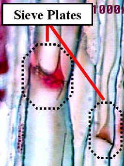
phloem. Sieve Tube Members:
more advance phloem conducting cells of angiosperms. Sieve tube members are short
and wide, and arranged end to end into sieve tubes. The sieve pores are large and
are concentrated along the end walls of adjacent sieve tube members. These
specializations allow solutes to move more rapidly in sieve tube members and sieve cells.
At maturity the nuclei in the sieve tube members disintegrates, the ribosomes
disappear, and the tonoplast (vacuole membrane) breaks down. Mitochondria and
plastids are still present. Sieve Tube Members are always associated with Companion
Cells which control the metabolism of the cells. These two cells are
connected by numerous plasmodesmata. The companion cells aid in the movement of
materials into and out of the sieve tube members. Sieve tube members also
contain P-protein, which stands for Phloem-protein. This protein is located along
the longitudinal walls of the cells. Some sieve tube members also contain a glucose
polymer called callose. Both P-protein and callose are responsible for sealing
wounds in the sieve tubes. |
 |
Back to Top of Page:
Back to Main
Plant Page