External
Morphology:
Internal Anatomy:
Shoot Apical Meristem
Primary
Stem Tissues
Dermal Tissue
Ground Tissue
Vascular Tissue
Secondary
Stem Tissues:
Vascular Cambium:
Cork Cambium:
Growth Rings:
Heartwood vs. Sapwood:
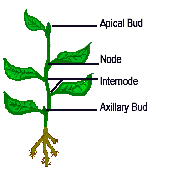
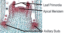
The node is the location of leaf attachment to the stem. The internode is that region of the stem between leaves. Apical meristems can be found within the apical and axillary buds. The apical meristem is responsible for formation of bud scales, leaves, and flowers or cones. The axillary buds will give rise to lateral branches.
Top of Page
Internal
Anatomy:
The internal anatomy of a plant will change as
the plant matures. The shoot tissues arise from the shoot apical meristem as dermal
tissue, ground tissue, and vascular tissue. Secondary meristems (vascular cambium
and cork cambium) then add girth to the plant by adding secondary xylem, phloem, and cork.
Top of
Page
Shoot Apical Meristem: the apical meristem includes the a group of dividing cells that give rise to three primary meristematic tissues, protoderm, ground meristem, and procambium. The outermost layer of cells is the protoderm which forms a single sheet of meristematic cells that give rise to the epidermis. This epidermis eventually covers all of the newly formed organs of the stem. The ground meristem forms the pith and cortex tissues of the stem and the mesophyll (middle leaf) tissue of the leaf. The procambium forms the primary xylem and phloem in both stems and leaves, as well and floral appendages.
Top of PagePrimary Stem Tissues
Dermal Tissue: The dermal tissue includes the epidermis of the stem and leaves with are covered by a cuticle. The cuticle is composed of a waxy material that keeps the underlying tissues from drying out. The epidermis of stems and leaves may contain hairlike extensions called trichomes. In leaves the epidermis may have stomata, small openings that are controlled by the action of guard cells. Guard cells contain chloroplasts, while epidermal cells and trichomes do not.
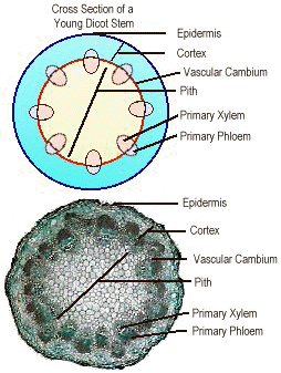
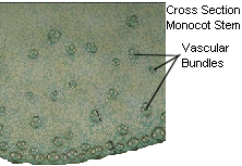
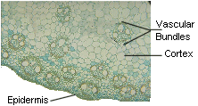
Top of PageGround Tissue: The ground tissue of a stem is divided into two regions, the cortex and the pith. The cortex is located to the outside and/or around the vascular bundles, while the pith is locate in the center of the stem. Both the cortex and pith are composed mainly of parenchyma cells. Support is given to the stem by collenchyma cells which may occur just under the epidermis in the cortex. This collenchyma cell layer may eventually develop into sclerenchyma. Monocots usually do not have a defined cortex and pith like that found in the Dicots. Monocots have their vascular bundles randomly scattered throughout the stem, while Dicots have their vascular bundles arranged in a ring.
Top of PageVascular Tissue: The primary vascular tissue forms longitudinal vascular bundles which run the length of the stem, and form veins in leaves. In monocot stems the vascular bundles are usually scattered, but in others (wheat, rice, oats) may form two rings. The scattered vascular bundles are interconnected and can form an intricate meshwork. In dicots and gymnosperms the vascular bundles form a ring around the pith. Vascular bundles branch off from the stem into the leaves of the plant. These branch points are called leaf traces, and occur at the base of the node. From the leaf trace, the vascular bundles subdivide to produce the veins of the leaf.
Top of PageSecondary Stem Tissues: In dicots and gymnosperms the majority of the plant tissue is composed of secondary vascular tissues.
Top of PageVascular Cambium: The vascular cambium is a layer of meristematic tissue that lies between the primary phloem and primary xylem. As the vascular cambium adds new xylem cells toward the inside of the stem and phloem cells toward the outside of the stem, the vascular bundles eventually coalesce to form a complete ring. The girth of the plant increases due to cell divisions perpendicular to the stem surface. The new cell walls are laid down in a radial direction, which increases the girth of the stem. There is more xylem formed than phloem. The new xylem forms concentric rings around the pith while the secondary phloem pushes the primary phloem outward. The old phloem becomes crushed and eventually sloughs off.
Top of Page
Cork Cambium: The cork cambium forms in the cortex of the stem. This meristem forms cork cells (phellem) to the outside and parenchyma cells (phelloderm) to the inside. The cork cambium, phellem, and phelloderm mad us the the periderm, or bark. This layer acts to protect the plant.
Top of Page
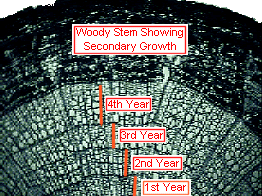
Growth Rings: growth rings are formed by the action of the vascular
cambium . During a single growing season in the temperature zone, large xylem cells
are produced during the early growing season in the spring. As the growing season
progresses into summer, the size of the new xylem cells decreases. This alternating
patter of large and small xylem cells produces the annual growth rings.
Top of Page
Heartwood vs.
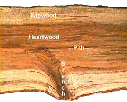
Sapwood: Heartwood is the darkened
central core of a woody stem. The heartwood contains the no longer
functional xylem which have been impregnated with tyloses. Sapwood is the lighter
outer portion of a woody stem. The sapwood contains functional xylem (those cells
involved in the transport of water an mineral nutrients).
Top of Page
